
Tips to Reduce False Positives in ADA Assays
Reducing false positives in ADA assays requires careful control of nonspecific binding, matrix interference, reagent quality, and assay cut points. Laboratories can improve accuracy by using well-characterized reagents, optimizing blocking and washing conditions, testing representative biological samples, and applying competitive inhibition during confirmatory testing. These methods help distinguish genuine anti-drug antibody responses from misleading signals caused by interfering proteins, pre-existing antibodies, or assay variability.
Proper immunogenicity testing also involves establishing statistically justified screening and confirmatory cut points, validating assay sensitivity and specificity, and monitoring performance across testing runs. Combining these practices helps laboratories reduce incorrect positive results while maintaining reliable antibody detection throughout drug development.
Understanding How False Positives Occur in ADA Assays
Identify Common Sources of Nonspecific Binding and Assay Interference
Nonspecific binding is one of the main sources of false-positive signals in ADA assays. It occurs when components in a biological sample interact with assay reagents, capture surfaces, detection labels, or the therapeutic drug without representing a genuine drug-specific antibody response. These interactions can increase background signals and cause samples to cross the screening cut point.
Several biological components can contribute to this problem. Rheumatoid factors, heterophilic antibodies, Fc receptors, complement components, and soluble drug targets may interfere with antibody detection, depending on the assay format. For example, rheumatoid factor can interact with immunoglobulin regions in assay reagents and produce misleading signals, particularly when the method relies on antibody recognition involving Fc regions.
Laboratory materials can also contribute to nonspecific binding. Microtiter plate surfaces, blocking proteins, detection reagents, and assay buffers may interact differently with individual serum samples. An unsuitable blocking reagent can reduce background in one assay configuration but introduce unexpected cross-reactivity in another. The FDA recommends evaluating blocking agents, wash conditions, detergents, and reagent interactions rather than assuming that a standard laboratory procedure will work equally well for every therapeutic product.
Understand Matrix Effects and Pre-Existing Antibodies
Matrix effects occur when components naturally present in serum or plasma influence the performance of an analytical method. Hemolysis, lipemia, bilirubin, endogenous proteins, concomitant medications, and disease-related changes in blood composition can alter assay signals. These effects may produce false-positive or false-negative results, depending on the sample composition and detection method.
Pre-existing antibodies require separate consideration. Some individuals may already have antibodies that recognize the therapeutic protein or share structural characteristics with it before treatment begins. Their presence does not automatically indicate a false-positive result. Instead, researchers must determine whether the baseline response represents genuine pre-existing reactivity, nonspecific interference, or an antibody response that increases following treatment.
This distinction becomes important when evaluating clinical samples over time. A patient who tests positive before receiving a therapeutic protein should not automatically be classified as having treatment-induced immunogenicity. Comparing baseline and post-treatment antibody responses, including changes in titer, can help establish whether treatment has generated or increased the response.
The EMA guideline discusses the importance of evaluating matrix-related interference, pre-existing immunity, and the clinical significance of antibody responses when assessing therapeutic proteins.
Evaluate Soluble Drug Target Interference
Soluble drug targets are another important source of misleading results, particularly in bridging immunoassays. These assays commonly use labeled versions of the therapeutic protein to capture and detect antibodies. A genuine ADA can bind both labeled drug molecules, forming a bridge that generates a measurable signal.
However, certain multivalent soluble target proteins can also interact with two labeled drug molecules and create a similar bridging effect. The resulting signal may resemble an antibody response even though the sample does not contain the intended drug-specific ADA.
This problem is not limited to screening assays. Depending on the assay design, soluble targets can interfere with confirmatory and neutralizing antibody measurements as well. Researchers should therefore investigate target concentrations, binding characteristics, and potential interference during assay development and validation.
Possible mitigation strategies include removing soluble targets, using suitable target-blocking reagents, or selecting an assay configuration that reduces the likelihood of unintended bridging. The Target interference research paper in The AAPS Journal provides further discussion of these mechanisms and their mitigation.
Practical Methods to Reduce False Positives in ADA Assays
Optimize Assay Design and Select High-Quality Reagents
A well-designed assay provides the foundation for reliable ADA detection. Laboratories should use purified, well-characterized therapeutic drug reagents with controlled aggregation, degradation, and stability. Aggregated or structurally altered proteins may expose binding regions that are not representative of the intended therapeutic molecule, increasing the possibility of nonspecific interactions.
Reagent labeling also requires careful optimization. Attaching biotin, enzymes, fluorophores, or other detection labels can change the structural properties of a therapeutic protein and affect antibody recognition. Excessive labeling or unsuitable coupling conditions may alter binding sites, reduce assay sensitivity, or increase background responses.
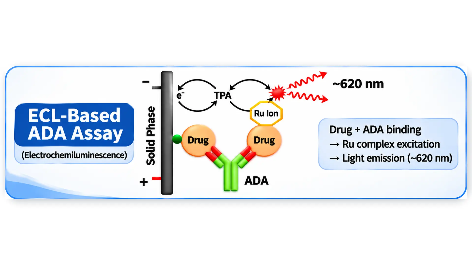
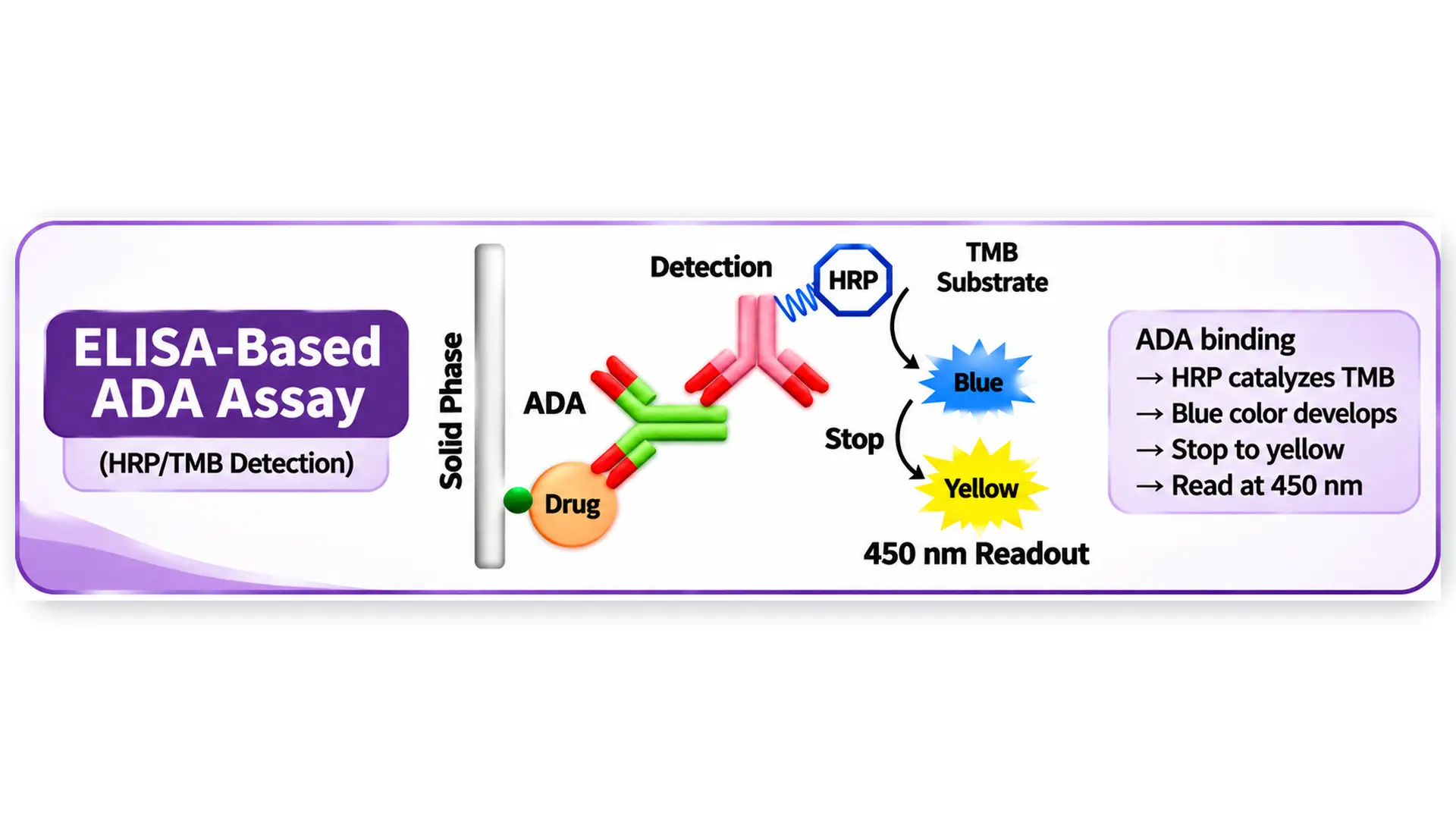
Assay format selection matters as well. Enzyme-linked immunosorbent assays (ELISA) and electrochemiluminescence (ECL) platforms are commonly used for ADA detection. ECL systems provide an instrument-based detection approach that can offer strong analytical sensitivity and useful signal-to-background performance. However, the detection platform alone does not guarantee specificity. Reagent selection, assay configuration, incubation conditions, and validation determine whether the method can distinguish genuine ADA responses from interference.
The United States Pharmacopeia addresses these considerations in its USP chapter on the design and validation of immunogenicity assays.
Improve Blocking Conditions and Control Background Signals
Blocking reagents help prevent unwanted interactions between biological samples and assay surfaces. Selecting an appropriate blocker can reduce nonspecific adsorption while preserving the binding activity required to detect genuine ADA responses.
Laboratories should evaluate multiple blocking conditions rather than relying on a single reagent without testing its suitability. The same principle applies to wash buffers, detergents, incubation periods, and washing sequences. Inadequate washing may leave unbound material that contributes to background signals, while excessively aggressive conditions may weaken genuine antibody interactions.
Testing unrelated proteins with comparable molecular size and charge can also help evaluate binding specificity. This provides additional information about whether a sample is interacting specifically with the therapeutic drug or responding to other assay components.
Use Sample Pretreatment Carefully
Sample pretreatment can improve assay performance when biological components or circulating therapeutic drugs interfere with antibody detection. However, the selected method should address the identified interference rather than introduce another source of analytical variation.
Acid dissociation is commonly used to disrupt circulating ADA-drug immune complexes and release antibodies that may otherwise remain unavailable for detection. This approach can improve drug tolerance, particularly when residual therapeutic protein concentrations are high. However, acid exposure may damage acid-sensitive antibodies or alter interactions involving soluble drug targets. Its suitability must therefore be established for the specific assay and therapeutic product.
Other approaches include affinity capture extraction, solid-phase extraction, and precipitation-based processing. These techniques can help separate interfering components from antibody-containing samples, although their effectiveness depends on the properties of the drug, antibody, and biological matrix.
Sample collection and storage procedures are equally important. Laboratories should evaluate sample stability under the storage, transportation, and freeze-thaw conditions expected during the study. Consistent collection tubes, anticoagulants, processing times, and storage procedures help prevent pre-analytical variation from affecting assay results.
Strengthen Specificity Through Competitive Inhibition
Competitive inhibition is a central part of confirmatory ADA testing. After a sample produces a positive screening result, researchers can repeat the analysis in the presence of an excess amount of unlabeled therapeutic drug.
If the detected antibody response is genuinely specific to the therapeutic protein, the unlabeled drug should compete with the assay reagents for antibody binding. This competition reduces the measured signal. If the signal does not decrease sufficiently under validated conditions, the original response may have resulted from nonspecific interactions or another form of assay interference.
The amount of inhibition required to classify a sample as confirmatory positive should be established through assay-specific validation. Applying an arbitrary percentage across different assay formats can produce incorrect classifications. Researchers should optimize the competitor concentration, incubation time, and detection conditions using appropriate negative and positive controls.
For example, consider a sample that crosses the screening cut point but fails to demonstrate the required drug-specific inhibition during confirmation. That sample should not automatically be reported as a confirmed ADA-positive result. Instead, its response should be interpreted according to the validated testing strategy, with further investigation if matrix effects or other interference remain suspected.
This additional testing step helps prevent nonspecific screening signals from being incorrectly included in immunogenicity assessments.
Strengthening ADA Assay Validation and Cut-Point Selection
Establish Statistically Appropriate Screening and Confirmatory Cut Points
Cut points determine whether a measured assay response is classified as positive or negative. Their selection directly affects the balance between sensitivity and specificity.
A screening cut point is generally designed to preserve sensitivity and identify samples that may contain ADA. It is therefore expected that some negative samples will produce screening signals above this threshold. FDA guidance describes an approximately 5% false-positive rate as a suitable target for initial screening. A subsequent confirmatory assay uses a more specific approach, with an approximately 1% false-positive rate generally recommended for its cut-point calculation.
These values should not be treated as arbitrary settings. Cut points must be established through appropriate statistical analysis using representative treatment-naïve samples. The sample population should reflect the intended study population because disease conditions and other biological differences can change background assay responses.
The FDA’s recommended screening cut-point approach generally uses around 50 treatment-naïve samples from the relevant subject population. Each sample should be tested by at least two analysts across at least three different days, providing six individual measurements. Statistical methods should account for the distribution of assay responses and justified outlier assessment.
A floating cut point may be appropriate when negative-control responses vary between assay runs or plates. It allows the threshold to account for changes in the assay’s background response while maintaining the intended statistical performance. Fixed cut points may be less suitable when the mean negative-control response changes during routine testing.
The FDA guidance provides detailed recommendations for screening, confirmatory, titration, and neutralization assay development and validation.
Use Representative Negative and Positive Controls
Appropriate controls help determine whether an assay is performing as expected. Negative controls should reflect the biological matrix of the study population and produce responses close to, but below, the established screening cut point. A control that consistently generates an extremely low signal may not reveal small increases in background response.
Low-positive controls are equally important. They help demonstrate that the assay can detect antibody responses near its sensitivity limit across different runs. If a low-positive control begins producing inconsistent results, the laboratory may need to investigate reagent degradation, changes in instrument performance, or variations in assay execution.
Positive and negative controls should undergo sample preparation and storage procedures comparable to those used for study samples. This helps ensure that control performance reflects the conditions under which actual clinical samples are tested.
Monitor Assay Performance and Reproducibility
Validation should not be considered the final step in controlling false positives. Assay performance can change over time because of reagent lot variation, instrument conditions, operator differences, and sample-related factors.
Laboratories should routinely monitor negative-control signals, low-positive control responses, confirmatory inhibition patterns, and assay acceptance criteria. Changes in these measurements may indicate increasing background noise or reduced analytical consistency.
Reproducibility should be evaluated within individual runs, between runs, across analysts, and over time. Selectivity testing should also include representative individual matrices, including samples with hemolysis, lipemia, and relevant disease-related characteristics. When the study population changes, previously established selectivity assumptions may need to be reassessed.
The AAPS harmonization paper discusses practical approaches to cut-point validation, system suitability controls, assay sensitivity, drug tolerance, matrix selectivity, and sample stability. The ABIRISK review also explains why assay interference should be considered when interpreting immunogenicity results.
Interpreting Confirmed ADA Results Correctly
Reducing false positives is not simply about obtaining fewer positive samples. Laboratories must also understand what a confirmed antibody response represents and whether it has any functional or clinical significance.
A confirmed binding ADA response indicates that antibodies in the sample specifically recognize the therapeutic drug. It does not necessarily mean that those antibodies interfere with the drug’s biological activity. Some antibodies bind to a therapeutic protein without affecting its function, while neutralizing antibodies (NAbs) can inhibit its biological activity.
Titration assays provide additional information by determining the highest sample dilution that still produces a response above the established assay threshold. This helps researchers characterize the relative strength of an antibody response, although an ADA titer should not be confused with an absolute antibody concentration.
Neutralizing antibody testing is a separate analytical step. Depending on the therapeutic drug’s mechanism of action, laboratories may use cell-based assays or non-cell-based competitive ligand-binding methods to determine whether detected antibodies inhibit biological activity.
Interpreting these results alongside pharmacokinetic, pharmacodynamic, safety, and efficacy data provides a more complete understanding of immunogenicity. A confirmed positive ADA result alone should not be treated as evidence of reduced treatment effectiveness or a clinically significant adverse response.
Conclusion
Reducing false positives in ADA assays requires a coordinated approach to assay development, reagent selection, sample handling, cut-point determination, and ongoing validation. Nonspecific binding, matrix interference, rheumatoid factors, pre-existing antibodies, and soluble drug targets can all influence measured responses. Identifying these sources early allows laboratories to select appropriate corrective measures without compromising the sensitivity required to detect genuine immune responses.
Competitive inhibition, representative treatment-naïve samples, validated statistical cut points, and suitable positive and negative controls provide a stronger foundation for reliable ADA testing. Sample pretreatment should also be selected according to the specific interference being addressed, with attention to its potential limitations.
As biotechnology continues to advance, analytical methods used to evaluate therapeutic proteins will remain an important part of pharmaceutical development. Reliable ADA assays help researchers distinguish genuine immune responses from misleading analytical signals, producing more dependable data for evaluating drug safety, efficacy, and clinical relevance.